
How Doppler Ultrasound Works and How to Set It Up
Doppler measures movement, not shape. Blood cells moving towards or away from the probe change the pitch of the echoes, and the machine turns that change into a colour, a spectrum and a velocity. Most Doppler mistakes come from three settings: the angle, the scale and the wall filter.
The Doppler shift in a blood vessel
Red blood cells scatter a small part of each ultrasound pulse. If they are moving towards the probe, the echo returns at a slightly higher frequency; if away, slightly lower. The difference is the Doppler shift:
- fD = 2 × f0 × v × cos θ ÷ c, where f0 is the transmitted frequency, v the blood velocity, θ the angle between the beam and the flow, and c is 1540 m/s.
- With a 4 MHz probe, blood at 1 m/s and a 60° angle, the shift is about 2.6 kHz — within human hearing, which is why Doppler can be played through a speaker.
- The machine measures fD and solves the same equation for v. Every velocity it reports therefore depends on the angle you tell it.
- By convention, flow towards the probe is shown above the baseline and, in colour, usually in red; flow away is below the baseline and usually blue. The colour bar on the screen shows which way round it is set.
Why the angle matters
The cos θ term is the weak point. At 90° cos θ is zero, so flow straight across the beam gives no Doppler signal at all. At steep angles a small error in the angle you set gives a large error in the velocity.
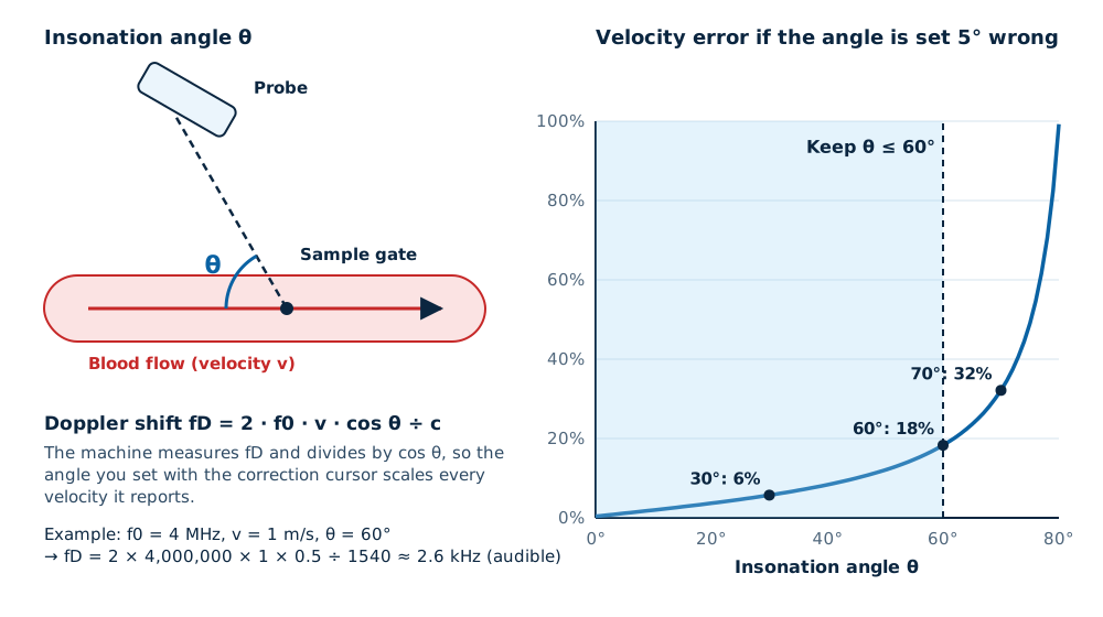
- Heel-toe the probe or steer the beam so the angle is 60° or less. The Society of Radiologists in Ultrasound consensus on carotid Doppler uses this limit.
- Align the angle-correction cursor parallel to the vessel wall, or to the jet direction in a stenosis.
- For follow-up studies, use a similar angle each time so that velocities can be compared.
- Colour and power Doppler images do not need angle correction to show that flow is present, but colour hue still depends on the angle.
Four Doppler modes and what each is for
| Mode | What it shows | Strengths | Limits |
|---|---|---|---|
| Colour Doppler | Average velocity and direction for every pixel inside a box, over the B-mode image | Finds vessels, shows direction, turbulence and jets | Slows the frame rate; aliases at high velocity; no exact peak velocity |
| Power Doppler | Strength of the Doppler signal, usually without direction | Most sensitive to slow and small-vessel flow; less angle-dependent | Usually no direction; very sensitive to probe and patient movement |
| Pulsed-wave (PW) spectral | Velocities over time from one small sample gate | Measures peak and mean velocity, resistance indices, at a chosen depth | Aliases above the Nyquist limit, which falls with depth |
| Continuous-wave (CW) spectral | All velocities along the whole beam line | Measures very high velocities, such as across a narrowed heart valve, without aliasing | Cannot tell at what depth the signal came from; needs a probe that supports it |
Read next Choosing a colour Doppler machine
PRF, the Nyquist limit and aliasing
Pulsed Doppler samples the moving blood once per pulse. It can only measure a Doppler shift up to half the pulse repetition frequency (PRF), the Nyquist limit. Above that, the signal wraps round and appears with the wrong direction: aliasing.
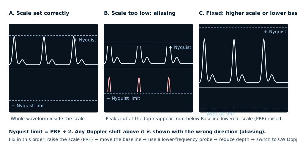
- The maximum measurable velocity is about c × PRF ÷ (4 × f0 × cos θ). With a 5 kHz PRF, a 4 MHz probe and a 60° angle, that is about 0.96 m/s.
- Deeper vessels force a lower PRF, because each pulse must return before the next is sent. Deep, fast jets are therefore the hardest to measure with PW Doppler.
- A lower transmitted frequency halves the Doppler shift for the same flow, which doubles the velocity you can measure before aliasing.
- In colour Doppler, aliasing shows as a sudden change from bright red to bright blue (or the reverse) in the middle of a vessel. Sometimes that is useful: it marks the fastest jet in a stenosis.
Wall filter, gain and the sample gate
Wall filter
Removes strong, low-frequency signals from moving vessel walls and tissue. Set too high, it also removes slow venous flow and end-diastolic flow, which distorts resistance indices. Use a low filter for veins and small organs.
Colour gain
Raise it until random colour speckles appear in the tissue, then lower it just enough for them to disappear. Too little gain hides slow flow; too much paints colour outside the vessel.
Sample gate (PW)
Place it in the centre of the lumen, smaller than the vessel, so that it samples the fast core of the flow and not the slow layers near the wall.
Colour box
Keep it as small and shallow as the question allows. A large, deep box lowers the frame rate and raises the thermal index.
Read next Knobology for B-mode settings
What is optimal: Doppler settings by task
| Task | Mode | Scale (PRF) | Wall filter | Notes |
|---|---|---|---|---|
| Carotid and leg arteries | Colour, then PW | Medium to high | Medium | Angle ≤60°, cursor along the wall, gate in mid-lumen |
| Leg veins (DVT) | Colour or power, PW for reflux | Low | Low | Compression first; augmentation to show flow |
| Umbilical artery in pregnancy | PW | Low to medium | Low | Use only when indicated; keep the thermal index low |
| Heart valves and jets | Colour, PW and CW | High; CW for fast jets | Medium to high | Align with the jet; CW needs a supporting probe |
| Ovarian, testicular or thyroid flow | Power or low-scale colour | Low | Low | Small box; compare both sides |
| Kidney stones (twinkle) | Colour | Medium | Default | Twinkling behind a stone helps confirm it |
Doppler and safety
Spectral Doppler concentrates energy at one place for some time, so it usually produces the highest thermal index of any mode. In pregnancy before 11–14 weeks, the WFUMB/ISUOG statement advises against routine pulsed Doppler; when it is needed, keep the displayed TI at 1.0 or below and the exposure short. Find the vessel in B-mode or colour first, switch to spectral Doppler only to measure, and freeze promptly.
Read next MI and TI: what the numbers mean
Doppler features to check before you buy
- Which probes support PW, colour, power and CW Doppler — CW in particular is often limited to phased-array probes.
- Colour sensitivity to slow flow on your own patients, at the depth you need.
- High-PRF mode for fast flow at depth, and automatic angle and gate tools if you scan vessels often.
- Measurement packages for the exams you report: vascular, obstetric or cardiac.
- Which Doppler modes are standard and which are licensed options on the quotation.
Read next Echo machines and CW DopplerHow ultrasound works (B-mode)
Related guides
Ultrasound Machine supply near you
Written quotes for clinics across Malaysia, handled from Batu Caves, Kota Kinabalu and Kuching.
Sources and further reading
- Boote EJ. AAPM/RSNA physics tutorial: Doppler US techniques. RadioGraphics 2003;23:1315–27
- Oglat AA et al. A review of medical Doppler ultrasonography of blood flow. J Med Ultrasound 2018
- Grant EG et al. Carotid artery stenosis: grayscale and Doppler US diagnosis — SRU consensus conference. Radiology 2003
- BMUS — Guidelines for the safe use of diagnostic ultrasound equipment (2009)
- AIUM — Recommended maximum scanning times for displayed thermal index values
Frequently asked questions
Why should the Doppler angle be 60 degrees or less?
What causes aliasing on Doppler ultrasound?
What is the difference between PW and CW Doppler?
Is power Doppler better than colour Doppler?
Why does flow disappear when the vessel is at 90 degrees to the beam?
Get a written quotation
Tell us your clinic or lab type, daily workload and location. We match suitable models and send a written quotation that states who handles delivery, installation and training, and what each costs.
010-905 2891 · Mon–Sat, 9:00 am – 6:00 pm