
How Ultrasound Works: From Piezoelectric Crystal to Image
An ultrasound scanner is a very fast sonar. The probe sends a short burst of sound, times every echo that comes back, and draws each one at the depth its delay implies. Here is how the probe, the gel, the timing and the choice of frequency fit together, and how to set them for each exam.
The short version
Diagnostic ultrasound uses sound far above human hearing, roughly 1 to 20 MHz, and it does not use ionising radiation. Each image is built line by line, many times a second:
- The probe converts an electrical pulse into a burst of sound a few cycles long.
- Gel carries that sound across the skin; without it almost all of it would bounce off the air gap.
- At every boundary between tissues, a small part of the sound reflects back as an echo.
- The probe turns each returning echo into a voltage. The machine converts the delay into depth and the strength into brightness.
- Repeating this along a hundred or more lines across the probe makes one frame. Repeating the frame gives a live, moving image.
Step 1: the piezoelectric effect in the probe
A piezoelectric material changes shape when a voltage is applied and produces a voltage when it is squeezed. Ultrasound probes use this both ways: the same elements transmit the pulse and receive the echoes.
Elements
Modern probes contain an array of small piezoelectric elements, usually a ceramic or a ceramic composite. Firing them with slightly different delays focuses and steers the beam electronically, with no moving parts.
Backing block
Damping material behind the elements stops them ringing after each pulse. A shorter pulse means finer detail along the beam, which is called axial resolution.
Matching layer
One or more layers on the face of the elements ease the sound from the hard ceramic into soft tissue, so less energy is reflected inside the probe.
Lens
The rubbery front face focuses the beam in the thin slice direction (elevation). It is also the part most easily damaged by the wrong wipes or by a drop.
Probe types and footprintsStep 2: pulse-echo timing turns time into depth
The machine assumes that sound travels through soft tissue at 1540 metres per second. Because each echo makes a round trip, depth equals speed multiplied by the delay, divided by two. That works out at about 13 microseconds of delay for every centimetre of depth.
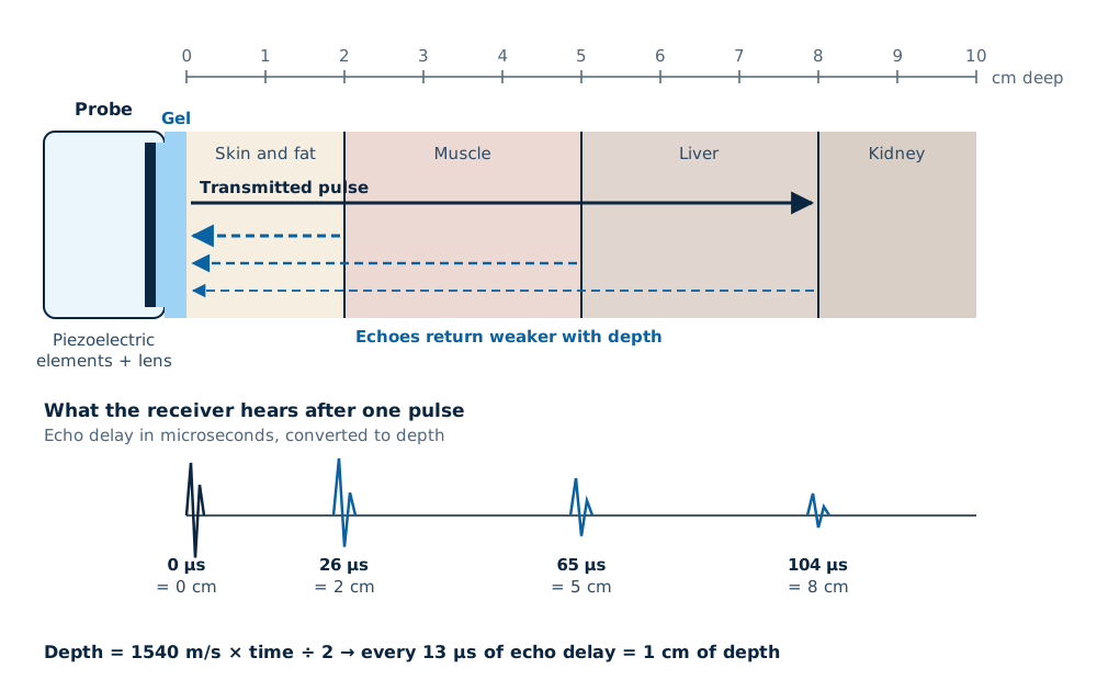
- The 1540 m/s figure is an average. Fat carries sound more slowly, so structures behind thick fat can be drawn slightly out of place; this is one source of artefacts.
- The probe cannot send the next pulse until the deepest echoes are back. Imaging to 15 cm limits the pulse rate to about 5,000 per second, and imaging to 20 cm to about 3,800.
- Frame rate is the pulse rate divided by the number of lines in the frame. More depth, a wider sector or extra focal zones all lower the frame rate.
Step 3: why echoes happen, and why gel is needed
Echoes come from changes in acoustic impedance, which is roughly a tissue’s density multiplied by its speed of sound. The bigger the change at a boundary, the stronger the echo. The fraction reflected is ((Z2 − Z1) ÷ (Z2 + Z1))².
| Boundary | Approximate impedances | Share of sound reflected | What you see |
|---|---|---|---|
| Fat to muscle | ≈1.4 and ≈1.7 | About 1% | A thin, faint line; most sound carries on deeper |
| Soft tissue to bone | ≈1.6 and ≈7.8 | About 40% | A bright surface with a shadow behind it |
| Soft tissue to air | ≈1.6 and ≈0.0004 | More than 99.9% | A bright line and nothing beyond it: why bowel gas and lung block the view |
| Probe to skin with gel | Gel is close to soft tissue | Very little | Sound enters the body; this is the whole point of gel |
Read next Ultrasound gel: types and handling
Step 4: attenuation and why deep echoes need help
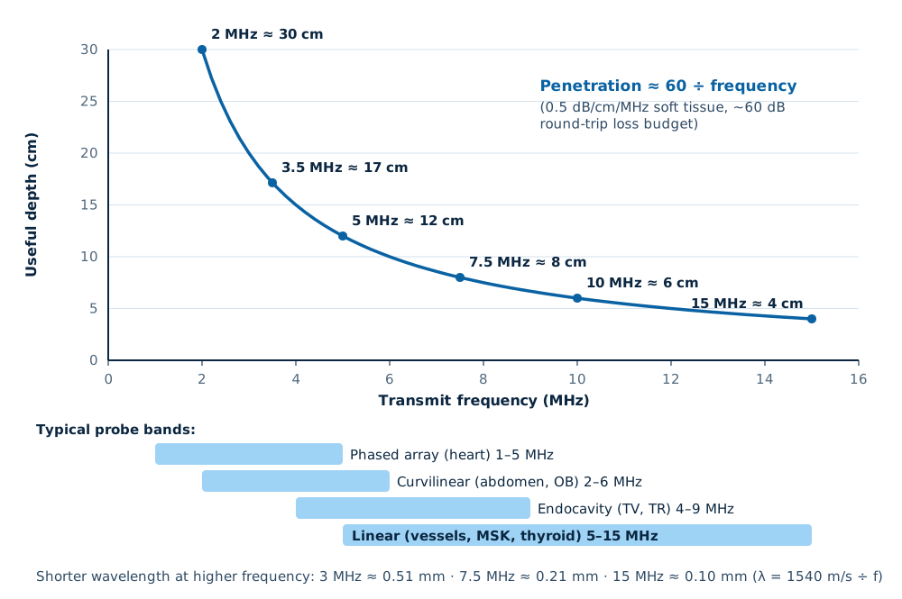
Sound loses energy as it travels, by absorption and scattering. In average soft tissue the loss is roughly 0.5 dB per centimetre for each megahertz of frequency, and it applies on the way down and on the way back. A 5 MHz pulse reflecting from 10 cm deep loses about 0.5 × 5 × 20 = 50 dB, a factor of about 100,000 in power.
- Time gain compensation (TGC) amplifies later echoes more than early ones, so the image looks even from top to bottom. Set the sliders so that a uniform organ such as the liver looks uniform.
- Fluid attenuates far less than tissue, so tissue behind a cyst or full bladder looks brighter (posterior enhancement).
- Bone, stones and gas stop most of the sound, leaving a dark shadow behind them.
Read next Knobology: gain, TGC, depth and focus
Frequency, depth and resolution: the trade-off
Higher frequencies have shorter wavelengths, so they show finer detail. They also lose energy faster, so they cannot reach as deep. Choosing a probe and frequency is choosing where on this curve to sit.
| Resolution | What sets it | How to improve it |
|---|---|---|
| Axial (along the beam) | Pulse length: about half the length of the pulse in tissue | Higher frequency, a well-damped probe |
| Lateral (across the image) | Beam width, narrowest at the focus | Put the focal zone at the structure of interest; use a higher frequency |
| Elevation (slice thickness) | The probe’s lens and, on some probes, a multi-row array | Choose a probe designed for fine slices; keep the target near the lens focus |
| Temporal (frame rate) | Depth, sector width, focal zones and line density | Reduce depth and sector width; use one focal zone for moving structures |
What is optimal: frequency and probe by exam
Use the highest frequency that still reaches the deepest structure you need to see, then set the depth so that structure fills most of the screen. Typical starting points:
| Exam | Probe | Typical frequency | Optimal set-up |
|---|---|---|---|
| Adult abdomen and kidneys | Curvilinear | 2–5 MHz | Depth 15–20 cm, harmonics on, focus at the organ |
| Large or deep patients | Curvilinear | Lowest setting, about 2 MHz | Harmonics on, reduce depth to what you need, single focus |
| Pregnancy after the first trimester | Curvilinear | 2–6 MHz | Obstetric preset, watch the thermal index |
| Early pregnancy and gynaecology | Endocavity | 4–9 MHz | Empty bladder, probe cover, depth to the pelvis only |
| Adult heart | Phased array | 1–5 MHz | Cardiac preset, narrow sector for a higher frame rate |
| Thyroid, breast, testis, skin | Linear | 7–15 MHz | Depth 3–5 cm, focus at the lesion |
| Carotid and leg veins | Linear | 5–10 MHz | Vascular preset, colour box small |
| Muscles, tendons and nerve blocks | Linear | 7–15 MHz or higher | Shallow depth, needle-visualisation mode if available |
Read next Abdominal ultrasound set-upRenal ultrasoundTransvaginal probes
Harmonics, compounding and other processing
Tissue harmonic imaging
Tissue distorts the pulse slightly as it travels, creating echoes at twice the transmitted frequency. Listening only to those reduces haze from the body wall and sharpens cyst edges. Most systems now have it on in their abdominal presets.
Spatial compounding
Combining frames steered at several angles smooths speckle and fills in edges, at some cost to frame rate.
Speckle reduction
Filters smooth the granular texture of the image. Too much makes the image look artificially soft.
What this means when you choose a system
- Look at each probe’s frequency range on its data sheet, not just the system brochure.
- Scan your heaviest typical patient at your deepest typical depth during the demonstration, and check the frame rate there.
- Check that harmonics work on the probes you will use most.
- Ask which processing features are standard and which are licensed options on the quotation.
- MDA registration applies to the system and to every probe (Medical Device Act 2012, Act 737).
Read next Choosing an ultrasound machineNext: how Doppler ultrasound worksCara ultrasound berfungsi (Bahasa Malaysia)
Related guides
Ultrasound Machine supply near you
Written quotes for clinics across Malaysia, handled from Batu Caves, Kota Kinabalu and Kuching.
Frequently asked questions
How does ultrasound work, in one sentence?
Why is gel needed for an ultrasound scan?
Why does a higher-frequency probe not see as deep?
Does ultrasound use radiation?
What is the speed of sound used by an ultrasound machine?
Get a written quotation
Tell us your clinic or lab type, daily workload and location. We match suitable models and send a written quotation that states who handles delivery, installation and training, and what each costs.
010-905 2891 · Mon–Sat, 9:00 am – 6:00 pm