
Echo Machine: How Cardiac Ultrasound Works and How to Choose One
An echo machine is an ultrasound system set up to image the heart: a small phased-array probe, high frame rates, M-mode, full Doppler including CW, and an ECG trace. How each of those works, the standard echo windows, the optimal specification for each type of practice, and what to test before you buy.
What makes an ultrasound an echo machine
Phased array probe
About 1–5 MHz with a small footprint that fits between the ribs.
M-mode
A motion trace along one line over time, used for wall and valve movement.
Full Doppler
Colour, PW and CW Doppler — CW is needed for high velocities across valves.
ECG trace
Timing images and measurements to the cardiac cycle.
Cardiac calculations
Measurement and report packages for chambers, function and valves.
How an echo machine images a moving heart
The heart is deep, behind ribs and lung, and moves fast. Each feature of an echo machine solves one of those problems.
Steering from a small footprint
A phased-array probe fires all its elements with tiny delays to swing the beam through a fan, so the whole image comes through one gap between the ribs.
How probes steer the beamFrame rate
Valves open and close in a fraction of a second. A narrow sector, only as much depth as needed and a single focal zone keep the frame rate high enough to see them.
M-mode
Samples one line thousands of times a second and plots it against time, giving the most precise timing of wall and valve movement.
Doppler and pressure
Velocities across valves are converted to pressure differences with the simplified Bernoulli equation: gradient in mmHg ≈ 4 × velocity² (m/s). A 4 m/s jet means about 64 mmHg. Jets that fast need CW Doppler, because PW would alias.
How Doppler works, and aliasingECG
Times each frame and measurement to the cardiac cycle, so end-diastolic and end-systolic measurements are taken at the right moment.
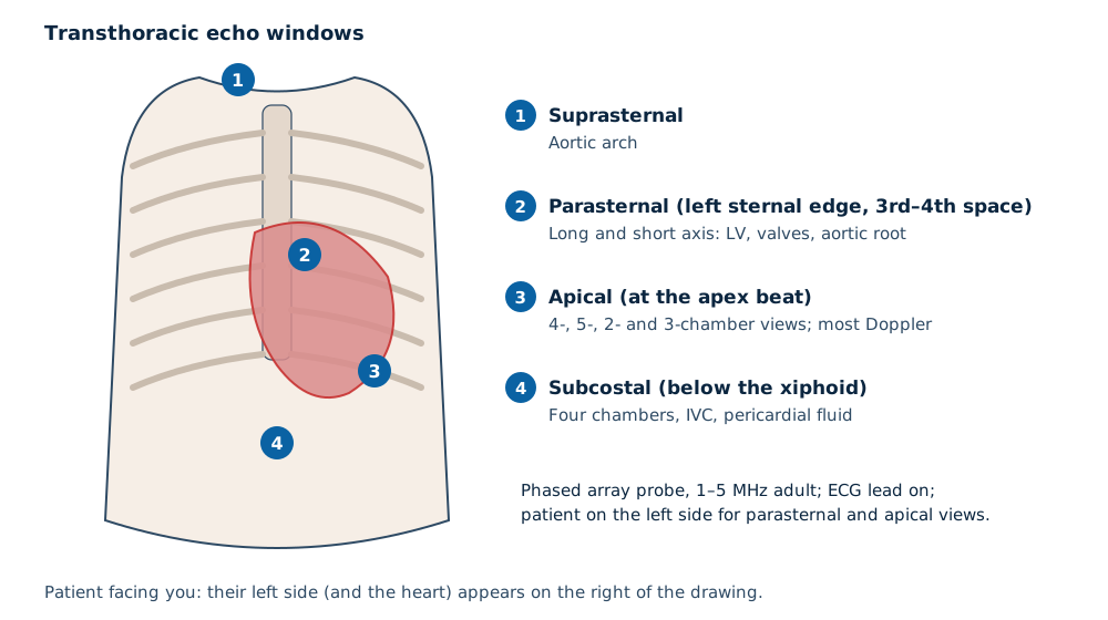
The standard echo windows
A transthoracic study combines views from four windows. Each gives a different line of sight through the heart.
Dedicated echo system or shared general system
| Dedicated cardiac system | General system with cardiac option | |
|---|---|---|
| Best for | Cardiology clinics and echo labs | Clinics doing occasional or focused cardiac scans |
| Probes | Adult and often paediatric phased array; TEE on some platforms | One phased array probe alongside convex and linear |
| Doppler | Strong CW and PW performance | Check that CW Doppler is included |
| Software | Full cardiac calculations and reporting | Basic cardiac measurements |
What is optimal: echo specification by practice
| Feature | Focused cardiac scans (POCUS) | Comprehensive adult echo | Paediatric echo |
|---|---|---|---|
| Probe | One phased array | Adult phased array | Small, higher-frequency phased arrays |
| CW Doppler | Useful | Essential | Essential |
| ECG input | Optional | Essential | Essential |
| Harmonics and high frame rate | Helpful | Essential | Essential |
| Cardiac calculations and reporting | Basic | Full package | Full package with paediatric references |
| Advanced options | Not needed | Strain, 3D and transoesophageal probes on some platforms | Depends on caseload |
| Connectivity | Export to the record | DICOM with structured reports | DICOM with structured reports |
Buying checklist
- Image quality on your own patients, including difficult windows.
- CW Doppler availability and which probes support it.
- ECG input and whether leads are included.
- Cardiac measurement and report packages, and DICOM export to your PACS.
- Paediatric or TEE probes if your practice needs them.
- Probe warranty, local service and MDA registration for the system and every probe.
Focused cardiac scans vs full echo
A focused cardiac scan at the bedside answers simple questions — pericardial effusion, gross ventricular function, volume status — and many portable systems handle it. A comprehensive echocardiogram needs the full Doppler set, ECG and cardiac calculations of a system built for it.
Read next POCUS equipmentDoppler modes explained
Related guides
Ultrasound Machine supply near you
Written quotes for clinics across Malaysia, handled from Batu Caves, Kota Kinabalu and Kuching.
Sources and further reading
- American Society of Echocardiography — Guidelines for performing a comprehensive transthoracic echocardiographic examination in adults (2019)
- Mitchell C et al. Comprehensive transthoracic echocardiographic examination in adults. J Am Soc Echocardiogr 2019
- Boote EJ. AAPM/RSNA physics tutorial: Doppler US techniques. RadioGraphics 2003
- BMUS — Guidelines for the safe use of diagnostic ultrasound equipment (2009)
Frequently asked questions
What is an echo machine?
Can a general ultrasound machine do echo?
Do paediatric echo studies need a different probe?
What is the difference between an echo machine and an ECG machine?
Why does an echo machine need CW Doppler?
Get a written quotation
Tell us your clinic or lab type, daily workload and location. We match suitable models and send a written quotation that states who handles delivery, installation and training, and what each costs.
010-905 2891 · Mon–Sat, 9:00 am – 6:00 pm