
Renal (Kidney) Ultrasound: Probe, Settings, Views and Measurements
A kidney scan is one of the most useful things a clinic ultrasound can do: it shows whether the kidneys are blocked, small, scarred or carrying stones and cysts, without radiation or contrast. How the kidney looks and why, the optimal probe and settings, the standard views and measurements, and how hydronephrosis is graded.
What a renal scan answers
- Is there hydronephrosis — dilatation of the collecting system that may mean obstruction?
- Are both kidneys present, in the usual place and of similar size?
- Are the kidneys small and bright, a pattern seen in long-standing kidney disease?
- Are there stones, simple cysts, or a solid mass that needs further imaging?
- How much urine is in the bladder before and after voiding, and is the bladder wall normal?
How the kidney looks, and why
Each layer of the kidney has a different structure, so it reflects sound differently. Knowing why helps you recognise what is abnormal.
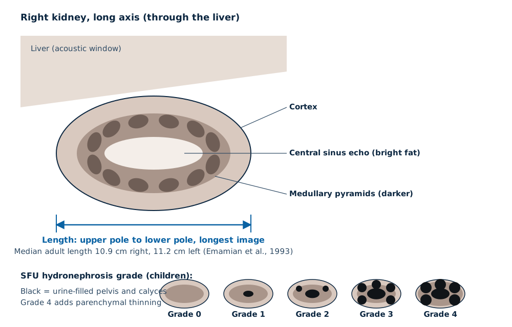
Cortex
Mid-grey. In adults it is normally no brighter than the neighbouring liver or spleen; a cortex brighter than the liver is a sign of medical renal disease.
Medullary pyramids
Darker triangles inside the cortex, because they contain more fluid and fewer reflecting interfaces. They are especially prominent in infants.
Central sinus
Bright, because fat and vessels around the collecting system create many strong interfaces.
Urine
Black, because fluid has no internal reflectors. A dilated pelvis or calyx shows as black branching spaces in the bright sinus, with brighter tissue behind (posterior enhancement).
Stones
Much stiffer than tissue, so they reflect strongly and leave a dark acoustic shadow. On colour Doppler many stones show a twinkling artefact behind them.
What is optimal: probe and settings
| Setting | Optimal starting point | Why |
|---|---|---|
| Probe | Curvilinear, about 2–5 MHz for adults; micro-convex or linear for children and thin adults | Penetration to the kidneys, which lie deep in the flank |
| Preset | Renal or abdominal | Sets dynamic range, persistence and harmonics for solid organs |
| Depth | Kidney fills about two-thirds of the screen | Larger image, higher frame rate |
| Focus | At the level of the kidney or the structure of interest | Narrowest beam where you are looking |
| Harmonics | On | Cleaner cyst walls and sharper stone shadows |
| Colour Doppler | Low scale for perfusion; colour over stones for twinkle; colour over the bladder base for ureteric jets | Each answers a different question |
| Gain | Liver and kidney cortex shown as mid-grey on the same image | Lets you compare cortical brightness fairly |
Read next Why frequency, depth and focus matter
Positions, windows and standard views
Right kidney
Patient supine or turned onto the left side. Scan through the liver at the right flank or anterior axillary line, with the breath held in deep inspiration to bring the kidney down.
Left kidney
Patient turned onto the right side. Scan further back, in the posterior axillary line, through the spleen; bowel gas often blocks an anterior approach.
Prone
Useful in children and when ribs or gas block the flank views; the kidneys lie just beneath the back muscles.
Views to record
Long axis through the longest length, then transverse at the upper pole, hilum and lower pole for each kidney, plus the bladder in two planes.
Measurements
- Kidney length: pole to pole on the longest long-axis image. In a study of 665 adult volunteers, median length was 11.2 cm on the left and 10.9 cm on the right; length correlated with height and decreased with age (Emamian et al., 1993).
- Compare the two sides. A kidney that is clearly smaller than its partner, or smaller than the patient’s earlier scans, needs explaining.
- Parenchymal thickness at mid-pole helps judge chronic scarring and thinning from long-standing obstruction.
- Bladder volume and post-void residual: length × width × height × a shape correction factor, calculated by most systems’ bladder package.
Read next Bladder scanners and post-void residual
Hydronephrosis: how it is graded
Grading describes how far the black, urine-filled collecting system spreads. It is a description, not a diagnosis of obstruction: a full bladder, a roomy extrarenal pelvis or pregnancy can all dilate a normal system.
- For antenatal and postnatal urinary tract dilatation, many units now use the UTD classification, which combines the anteroposterior pelvic diameter with calyces, parenchyma, ureters and bladder (Nguyen et al., 2014).
- In adults, reports commonly describe hydronephrosis as mild, moderate or severe, together with the cause if seen.
- If the bladder is very full, rescan the kidneys after the patient has voided; mild dilatation often resolves.
- Parapelvic cysts can mimic hydronephrosis; unlike a dilated pelvis, they do not connect with each other.
| SFU grade | Ultrasound appearance |
|---|---|
| 0 | No dilatation; the central sinus is intact |
| 1 | Renal pelvis visible, no calyces seen |
| 2 | Pelvis dilated, a few calyces visible |
| 3 | Nearly all calyces dilated, parenchyma normal |
| 4 | As grade 3, with thinning of the parenchyma |
Kidney stones and cysts on ultrasound
Stones
Look for a bright focus with a clean shadow, and twinkle on colour Doppler. Small stones and stones in the middle ureter are easily missed; a normal scan does not exclude a small stone.
Simple cysts
Round, black, thin-walled and with posterior enhancement. Cysts with septa, solid parts or internal flow need further imaging.
Ureteric jets
Colour Doppler over the bladder base shows urine squirting in from each ureter. A jet from both sides argues against complete obstruction.
Equipment checklist for renal scanning
- A curvilinear probe with good penetration on heavy patients, and harmonics on that probe.
- Colour Doppler sensitive enough for ureteric jets and stone twinkle.
- Measurement and bladder-volume packages included, not licensed extras you did not expect.
- DICOM export so that kidney lengths can be compared between visits.
- MDA registration for the system and every probe (Act 737).
Read next Ultrasound for GP clinics
Related guides
Ultrasound Machine supply near you
Written quotes for clinics across Malaysia, handled from Batu Caves, Kota Kinabalu and Kuching.
Sources and further reading
- Emamian SA et al. Kidney dimensions at sonography in 665 adult volunteers. AJR 1993;160:83–6
- Fernbach SK et al. Ultrasound grading of hydronephrosis: the Society for Fetal Urology system. Pediatr Radiol 1993
- Nguyen HT et al. Multidisciplinary consensus on the classification of urinary tract dilation (UTD). J Pediatr Urol 2014
- AIUM — Practice parameters for ultrasound examinations
Frequently asked questions
What is the normal size of a kidney on ultrasound?
Which probe is used for a kidney ultrasound?
Do I need a full bladder for a kidney ultrasound?
Can ultrasound see kidney stones?
What does hydronephrosis look like on ultrasound?
Get a written quotation
Tell us your clinic or lab type, daily workload and location. We match suitable models and send a written quotation that states who handles delivery, installation and training, and what each costs.
010-905 2891 · Mon–Sat, 9:00 am – 6:00 pm