
Transvaginal Ultrasound Probe: How It Works, Uses and Reprocessing
A transvaginal (endocavity) probe puts the transducer a few centimetres from the uterus and ovaries, so it can use a higher frequency and show far more detail than a scan through the abdomen. It also touches mucous membrane, which makes cleaning and high-level disinfection part of every scan.
What a transvaginal probe is
An endocavity probe has a small curved array at the tip of a long, slim handle. It is used transvaginally, and with the right cover also transrectally. Typical frequencies are about 4–9 MHz, and the array produces a wide fan-shaped field from the tip.
Tip array
A micro-convex array on the end of the shaft, often angled slightly, giving a wide sector close to the probe.
Handle and orientation mark
A ridge or light shows which side of the image corresponds to which side of the probe, which matters when you rotate between sagittal and coronal planes.
Biopsy guide
Many models accept a needle guide for procedures such as egg retrieval; check which guides fit the model on your quotation.
Why it shows more detail
Detail in ultrasound is limited mainly by frequency, and frequency is limited by depth. Placing the probe next to the pelvic organs shortens the sound path from perhaps 10 cm or more through the abdominal wall and full bladder to a few centimetres, so a higher frequency can be used.
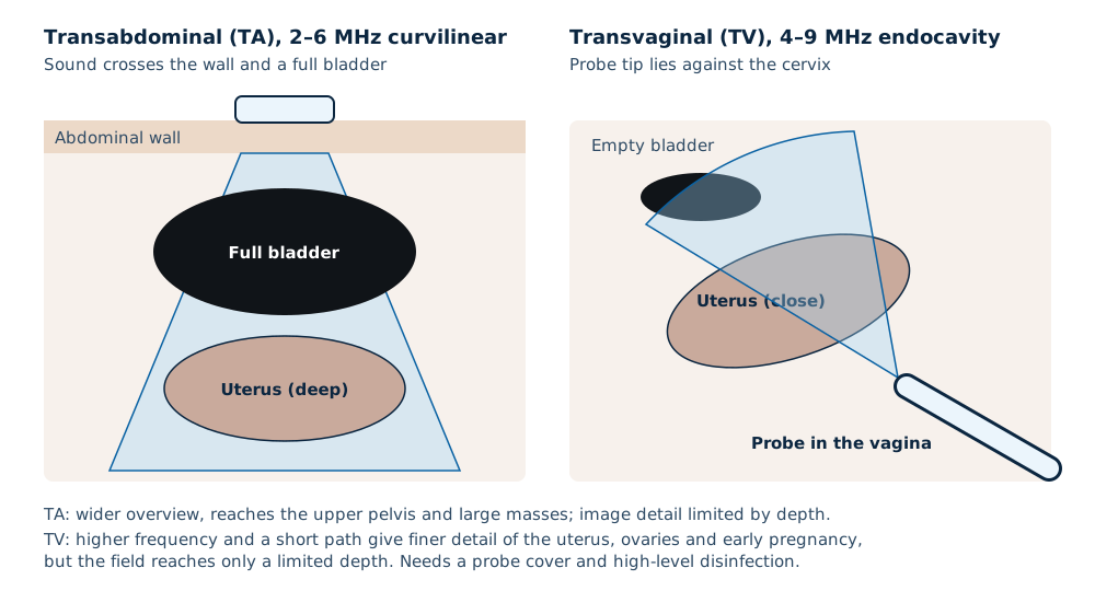
- No full bladder is needed, and bowel gas is rarely in the way.
- The trade-off is reach: the field of view ends a short distance from the tip, so large masses and the upper pelvis may need a transabdominal view.
- Higher frequency also means higher sensitivity to probe position — small movements change the image a lot.
What it is used for
| Use | What the scan looks for |
|---|---|
| Early pregnancy | Location of the pregnancy, the gestational sac, yolk sac, embryo and heartbeat, and crown–rump length for dating |
| Pregnancy of uncertain viability | Criteria applied on transvaginal scans, such as an embryo with crown–rump length of 7 mm or more and no heartbeat, or a mean sac diameter of 25 mm or more with no embryo, which are diagnostic of pregnancy failure (SRU consensus, 2013) |
| Gynaecology | Endometrial thickness and pattern, fibroids, ovarian cysts, adnexal masses, pelvic fluid |
| Fertility | Antral follicle count, follicle tracking during stimulation, endometrium before transfer |
| Other | IUCD position, cervical length in pregnancy when indicated, pelvic pain |
Read next Follicle tracking ultrasoundObstetric ultrasound machines
What is optimal: settings and technique
- Ask the patient to empty her bladder first; a full bladder pushes the organs away from the probe.
- Put gel inside the cover and on the outside of the cover; use sterile gel and a sterile cover where your protocol or a procedure requires it.
- Use the highest frequency that reaches the area of interest, and reduce the depth so the uterus or ovary fills the screen.
- Place the focal zone at the structure you are measuring.
- Sweep systematically: sagittal from side to side, then rotate 90° to coronal and sweep from top to bottom.
- Keep the thermal index low in early pregnancy and avoid routine pulsed Doppler before 11–14 weeks.
- Offer a chaperone, explain each step, and record consent and who was present according to your clinic policy.
Read next MI and TI in early pregnancyRoom set-up, privacy and chaperones
Covers and high-level disinfection
Under the Spaulding classification, a transvaginal probe is a semi-critical device because it touches mucous membranes. The WFUMB Safety Committee advises a new cover for every patient and high-level disinfection between patients, because covers can leak: older studies reported leaks in 0.9–2% of condoms and 8–81% of commercial probe covers.
- Remove and discard the cover.
- Clean off gel and debris with running water or a damp cloth and mild non-abrasive soap, then dry.
- High-level disinfect with a method the probe manufacturer approves for that model.
- Disinfect the handle and the cable nearest the handle too, which also get touched.
- Store the probe so it cannot be recontaminated, and fit a new cover for the next patient.
- Keep a record of each disinfection cycle where your method allows it.
| Method | Example contact time in the guideline | Notes |
|---|---|---|
| Glutaraldehyde 2.4–3.2% | 20 minutes at 20 °C | Strong odour; needs ventilation; largely replaced |
| Ortho-phthalaldehyde (OPA) | 5 minutes at 20 °C | Milder odour, no activation needed |
| Chlorine dioxide wipe systems | Seconds per wipe step | Three-step pre-clean, disinfect and rinse wipes |
| Hydrogen peroxide 7.5% | Per product instructions | Broad activity, including spores |
| Automated hydrogen peroxide mist | Per device cycle | Closed system that also treats the handle; needs the device |
Buying a transvaginal probe
- It must be compatible with your system platform and software version; probes are not interchangeable between brands.
- Compare the frequency range and the field-of-view angle on the data sheet.
- Check which needle guides fit, if you do procedures.
- Ask for the manufacturer’s list of approved disinfectants and covers, and check it matches what your clinic uses.
- Probe warranty terms, and how drops or cable damage are treated.
- Diagnostic ultrasound systems and their transducers are medical devices. Under the Medical Device Act 2012 (Act 737), a medical device must be registered with the Medical Device Authority (MDA) before it is imported, exported or placed on the Malaysian market — ask for the registration number and check it on the MDA’s public register. Ultrasound uses sound, not ionising radiation, so unlike X-ray it does not need a licence under the Atomic Energy Licensing Act 1984.
Related guides
Ultrasound Machine supply near you
Written quotes for clinics across Malaysia, handled from Batu Caves, Kota Kinabalu and Kuching.
Sources and further reading
- WFUMB Safety Committee — Guidelines for cleaning transvaginal ultrasound transducers between patients (Ultrasound Med Biol 2017)
- AIUM — Guidelines for cleaning and preparing external- and internal-use ultrasound transducers, 2025 revision
- Doubilet PM et al. Diagnostic criteria for nonviable pregnancy early in the first trimester. N Engl J Med 2013
- ISUOG — Practice guidelines
Frequently asked questions
What frequency is a transvaginal ultrasound probe?
Why is a transvaginal scan clearer than an abdominal scan?
Does a transvaginal probe need high-level disinfection?
Can the same probe be used transrectally?
Should the bladder be full for a transvaginal scan?
How often should a transvaginal probe cover be changed?
Get a written quotation
Tell us your clinic or lab type, daily workload and location. We match suitable models and send a written quotation that states who handles delivery, installation and training, and what each costs.
010-905 2891 · Mon–Sat, 9:00 am – 6:00 pm